New concepts for mitral valve imaging
Abstract
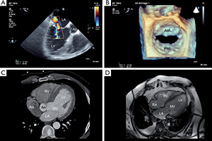
The high complexity of the mitral valve (MV) anatomy and function is not yet fully understood. Studying especially the dynamic movement and interaction of MV components to describe MV physiology during the cardiac cycle remains a challenge. Imaging is the key to assessing details of MV disease and to studying the lesion and dysfunction of MV according to Carpentier. With the advances of computational geometrical and biomechanical MV models, improved quantification and characterization of the MV has been realized.
Geometrical models can be divided into rigid and dynamic models. Both models are based on reconstruction techniques of echocardiographic or computed tomographic data sets. They allow detailed analysis of MV morphology and dynamics throughout the cardiac cycle. Biomechanical models aim to simulate the biomechanics of MV to allow for examination and analysis of the MV structure with blood flow. Two categories of biomechanical MV models can be distinguished: structural models and fluid-structure interaction (FSI) models.
The complex structure and dynamics of MV apparatus throughout the cardiac cycle can be analyzed with different types of computational models. These represent substantial progress in the diagnosis of structural heart disease since MV morphology and dynamics can be studied in unprecedented detail. It is conceivable that MV modeling will contribute significantly to the understanding of the MV.
Cover